


Diseases of the musculoskeletal system are almost always accompanied by dysfunction of the organs, which causes a lot of discomfort to the patient.
An important symptom of musculoskeletal pathologies is pain. Joint injuries are particularly unpleasant.
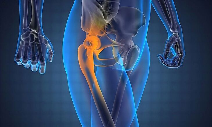
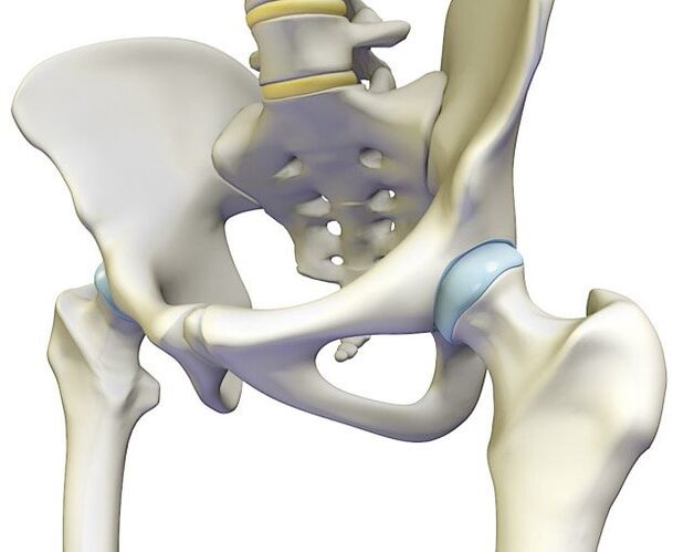
The hips are the largest of them. Pain in the event of defeat can be localized around it and can be transmitted to different anatomical structures: the pelvic, lower back or thigh organs.
General classification of causes
The etiology of hip pain is varied.
In medicine, the following causes of arthralgia are conditionally distinguished:
- Inflammatory and infectious processes in the joint and surrounding tissues.
- Degenerative diseases of the musculoskeletal system.
- Injuries.
- Neoplasms of bones and soft tissues.
There are other specific causes of arthralgia:
- Piriformis syndrome. It is associated with prolonged convulsions.
- Femoral head necrosis (GBC). It is most often a complication of another pathology of TBS.
- Legg-Calve-Perthes disease. Osteochondropathy of the GBC.
- Autopsy of osteochondrosis. It can be called Koenig's disease in various sources.
- Diabetic osteoarthropathy. A complication of diabetes.
- Pseudogout. Also chondrocalcinosis.
- Periodic hydrarthrosis is an overproduction of synovial fluid.
- Synovial chondromatosis (Lotsch syndrome).
In addition, the legs in the hip area of pregnant women often hurt.
During this period, complex hormonal changes occur, with the growing uterus displacing adjacent organs and straining the ligament apparatus of the hip joint. In addition, weight gain increases the load on the legs. Failure to follow dietary recommendations can lead to calcium deficiency in the pregnant woman, which upsets the mineral balance and disrupts the structure of the bones and joints.
Causes of pain
The incidence of arthralgia increases with age.Symptoms of TBS (hip joint) occur in up to 10% of children and 50% of the elderly. Most often, women suffer from this pathology. This is due to age-related hormonal changes after menopause.

Why does my hip hurt? There is no definite answer to this question, as the list of reasons is quite long.
The main factors that cause arthralgia of the hip joint are:
- Pathological process inside the muscle tape device. Most often, this is due to a direct mechanical effect: bruising of the joint with subsequent inflammation of the components.
- Anatomical changes in the joint. They can be congenital or post-traumatic (dislocations, fractures).
- Pathology of other systems. Inflammation of the organs of the MT (pelvis) can spread to the pelvic bones. Neurological disorders manifest in pain of any localization. Metabolic disorders cause mineral imbalance. The bone-tape connection weakens and the risk of injury increases.
Inflammatory and infectious processes in the joints and surrounding tissues
The most common cause of joint pain of any localization is musculoskeletal joint suppuration.
Inflammation of the hip joint can be divided into:
- Primary. It is caused by pathogens penetrating directly into the joint: hitting with a sharp or blunt object, forming a wound.
- Secondary. TBS infection occurs from a distant foci of inflammation: by contact or hematogenously.
Arthritis TBS
It is most common in elderly patients.Aching pain in the hip joint, exacerbated by walking, radiates to the groin, perineum and thigh. The patient has difficulty getting up from the chair or climbing the stairs without assistance. The feeling of discomfort is worse in the morning.
Therapy includes taking anti-inflammatory drugs and delivering glucocorticoids to the intra-articular burst. If necessary, drain its cavity.
Rheumatoid arthritis (RA)
It is a chronic systemic connective tissue disease similar to polyarthritis.The essence of this pathological process is inflammation of the synovium, cartilage, and joint capsule. The cause is a malfunction of the immune system. It is characterized by polyarthralgia, stiffness of morning movements, high fever possible.
The shoulder and hip joints are extremely rarely affected, with pain occurring only in the late stages of RA, a few years after the onset of the disease.
Acute septic arthritis
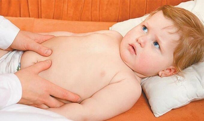
It is an infectious childhood disease, with 70% of cases occurring in infants under 4 years of age. The pathogen is usually Staphylococcus aureus. The child refuses to walk because of severe acute pain in the hip joint and groin while moving. It is characterized by high fever and increased irritability.
Treatment includes removal of the effusion from the joint cavity and antibiotic therapy.
The risk of developing osteomyelitis and sepsis is high.
Tuberculous coxitis or arthritis
Most often, pediatricians face this disease. In young children, the immune system is poorly developed, leading to the possibility of infection.
This disease is characterized by slow progression. Initially, the child gets tired very quickly, his activity decreases, he stops running. Gradual atrophy of the thigh muscles occurs. Movements are impeded. The pain in the child’s hip joint is intense, the limb will be longer than a healthy one.
When pus melts the joint membrane, the secretion spreads along the muscles and tendons, forming phlegmons and fistulas.
Conservative treatment is given in the absence of complications.
Tendovaginitis in the hip joint
This pathology is an inflammation of the muscle tendon and vagina. Causes prolonged overload or foot injury.
Main complaints: thigh joint pain during movement, the swelling swells, gait change - lameness becomes noticeable.
Treatment - medication: intra-articular injections of anti-inflammatory drugs, corticosteroids.
Bursitis
Of all the synovial sac, the acetabulum bursa was the most commonly inflamed.It partially covers the femur. In hip bursitis, the pain radiates to the thigh and gluteal region. The patient is unable to lie on the affected side: the pressure in the synovial sac increases and the pain increases.
If the bursitis does not cause complications, treatment consists of unloading the lower limb with a stick or crutch.
Medications: Analgesics and corticosteroids.
Idiopathic ankylosing spondylitis
It is a chronic inflammation of the elements of the spine and sacroiliac joints.
The disease is dangerous because of its complications that reduce living standards and lead to disability.
If you notice such a problem, contact a professional immediately to determine the appropriate treatment.
The etiology is not entirely clear. According to modern medicine, the leading cause is hereditary predisposition. People under the age of 30 get sick the most.
Symptoms of ankylosing spondylitis:
- Elevated body temperature, fever.
- Poisoning syndrome: general malaise, weakness, loss of appetite, weight loss, sleep disturbance.
- Constant dull pain in the hip joint and at the level of the sacrum and buttocks that spreads to the back of the thigh. They are usually bilateral and increase in intensity at night.
- Limited mobility in the lower back and hips. This symptom gradually passes to the upper parts of the spine along the entire back, including the neck. As a result, the patient is forced to assume the "applicant's pose. "
Rehabilitation therapy is based on special physiotherapy exercises for joint development.
Medications: NSAIDs to relieve pain and inflammation, corticosteroids.
Tendinitis
Athletes or people whose work involves hard physical work are prone to inflammation of the tendons. Characteristics of the manifestation: Aching pain occurs in the hip joint under heavy load. At rest, the discomfort is usually not observed.
It is recommended to reduce the load on the feet, in advanced cases - bed rest.
Medication: NSAIDs, topical analgesic gels, glucocorticosteroids, chondroprotectors.
Syphilis
In the late stages of the disease, the bones and joints are affected. Rubber formation is typical. Excessive pathological mineralization occurs. TBS is extremely rare.
Gumma - a node in the tissues that is formed during advanced syphilis and destroys the surrounding tissues. The process ends with the formation of rough scars.
Treatment is ineffective and the risk of developing complications in the form of osteomyelitis is high.
Fungal arthritis of the hips
This is due to the long-term use of antibiotics and pathologies of the immune system.
People living with HIV or AIDS are particularly susceptible to fungal arthritis.
Joint pain is constantly present, painful in nature.
Fungal lesions in the bones are characterized by a tendency to develop a fistula, and the duration and difficulty of treatment.
Therapy: systemic antimycotics.
Surgery is indicated.
Tumors of bones and soft tissues
The oncological diseases of the hip joint can be cancers of a distant organ or they can develop on their own.
- Benign tumors of the bone tissue - osteomas.
A formation foreign to the body grows, compressing nerves and blood vessels. The clinic is similar to piriformis syndrome.
- Malignant bone tumors - osteosarcomas.
The neoplasm grows rapidly, dying, disintegrating, spreading metastases throughout the body.Nocturnal pain in the hip joints is unbearable and does not go away even after taking NSAIDs or anesthesia.
- Mesenchymal tumors are made up of soft tissues. Benigns rarely recur and do not metastasize. The intensity of pain varies depending on the aggressiveness of the malignant cells.
Degenerative diseases of the joints
Coxarthrosis
Hip arthrosis is a chronic disease characterized by altered integrity of the joint surfaces due to a violation of metabolic processes. It develops very slowly over several years. Initially, cartilage tissue is affected, followed by bone tissue, followed by varus deformity of the joint and limb. Occurs at age 40.
Symptoms:
- The hip joint only hurts while walking.
- Stiffness of movements in the TBS.
- As the process progresses, a shortening of the length of the limb is observed.
- Weakness and atrophy of muscle mass.
- Lameness.
- You will hear a crack as you walk.
- In the case of a bilateral lesion, a "duck walk" occurs - a transfer from one foot to another.
Medications: NSAIDs, vasodilators, muscle relaxants, chondroprotectors, injection of hormonal drugs into the joint cavity.
Local effects: ointments, body lotions, compresses.
Surgery is in the final stages of the disease.
Osteochondrosis
Degenerative changes in the intervertebral discs damage the surrounding tissues.
Symptoms:
- Pain in the lower back that radiates to the hip joint and thigh.
- Suddenly, sharp and sharp. It begins in the lumbar region and in the buttocks, descending at the back of the foot.
- Unilateral localization of pain is more common.
- The patient is forced to lie down - lying on a healthy side.
- The sensitivity of the skin of the foot may have decreased.
The treatment is complex. Anti-inflammatory and analgesic drugs, moderate physical activity (swimming), physiotherapy after alleviation of the most acute phenomena.
In case of severe pain, anesthesia is recommended.
Injuries
Injury
It is characterized by moderate pain, its intensity increases during active movements. After injury to the hip joint, lameness first appears and resolves quickly.
At rest, the symptoms disappear.
In order to get rid of pain quickly in the case of a pelvic joint injury, a cold should be applied to the site of the injury: an ice pack or a frozen product.
hip dislocation
May:
- He was born. This is the result of pathologies of miscarriage or intrauterine development. The child has uneven tail wrinkles and shortening of the limb, possibly pinched nerve, which manifests itself in cramps. If the dislocation is not corrected in infancy, the child may later become disabled.
- Traumatic. Signs: Acute severe pain, complete cessation of joint function, massive edema, and extensive hematoma appear over the injured area. Without help, it becomes impossible for the patient to get out of the chair or bed.
In case of hip dislocation, go to the emergency department or hospital immediately.
fractures
The hip joint is made up of strong strong bones.
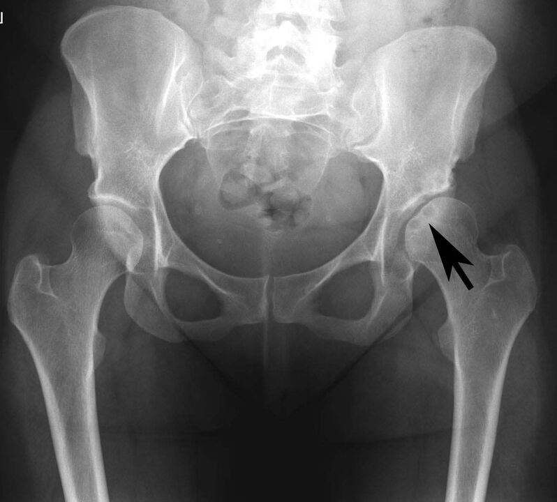
The most common diagnosis in this subgroup is fracture of the femoral neck. It is mainly recommended for women after 60 years.
The cause of such damage is the fall or collision of the TBS area.
The strongest pain is felt, the hip joint is stretched, abscess, movement is almost impossible in it. The upper part of the thigh swells, an extensive hematoma appears. The injured leg is shortened and the patient limps. A characteristic click is heard as you move.
In the event of a fracture, the surrounding tissues are damaged, resulting in a burning sensation. In the absence of treatment, an inflammatory process can begin here. If the nerve is pinched, you may experience numbness in your thighs.
The treatment is complex: surgical and medical.
Specific causes of arthralgia
Piriformis syndrome
With the localization of pathological processes in the area of the hip joint, the surrounding tissues are also affected. Prolonged spasmodic piriform muscle compresses the sciatic nerve and its blood vessels, causing a number of symptoms:
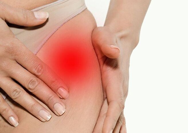
- Pain in the leg in the region of the hip joint. It goes to the buttocks and the lumbosacral joint.
- Increased discomfort when leaning on the affected foot.
- Compression of piriformis muscle.
- Sudden pain "lumbago" along the nerve.
Etiology: pelvic organ injuries, infectious and inflammatory diseases, vertebrogenic pathologies, muscle overexertion, long-term preservation of non-physiological posture.
Medications: NSAIDs, muscle relaxants, analgesics, circulatory enhancers, glucocorticoids.
After the relief of acute phenomena, rehabilitation measures may be prescribed: physiotherapy, massage, acupuncture.
Aseptic necrosis of the femoral head
It occurs in most young men. The etiology of the disease is ischemia of the upper thigh. Insufficient blood supply to the tissues results in oxygen starvation and the onset of necrosis (necrosis).
Clinical picture: the hip joint hurts and gives to the foot and perineum. It is not possible to rely on a damaged leg. After a few days, the nerve endings melt and the pain disappears. That's a terrible sign! There is a high risk of rapid development of osteomyelitis and sepsis in the case of necrosis of the deeper layers of the bone.
Treatment is surgery and medication.
Koenig's disease
Autopsy osteochondritis - detachment of a small area of necrotic cartilage from the bone and its protrusion into the joint cavity.
It is a rare disease. It is typical for men aged 15-35 years.
Patients complain of mild aching pain in the hip joint. The joint "gets stuck" during movement.
Treatment is conservative (duration 10-18 months) and surgical. During the surgical procedure, the exfoliating masses are removed, and the uniformity (comparability) of the joint surfaces is restored.
Diabetic osteoarthropathy
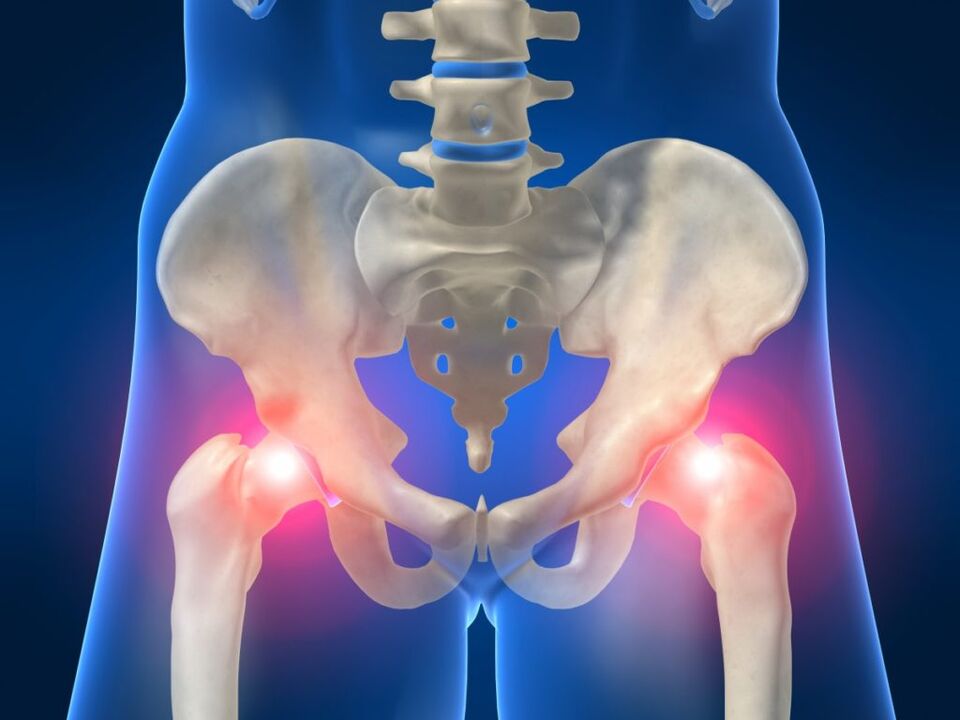
Violation of glucose metabolism leads to circulatory disorders and innervation of all organs. Changes in the hip joint are more often unilateral: they occur more often on the right side than on the left side. The immune response is reduced, which makes it easier for the body to infect.
Clinical picture:
- Swelling of the joint.
- The skin above it is cold to the touch.
There is no pain syndrome in diabetic osteoarthropathy!
Treatment consists of careful monitoring of blood sugar levels and timely administration of insulin.
pseudogout
This pathology is the deposition of calcium salts in the articular cartilage.
Doctors associate it with endocrine pathologies: hyperparathyroidism, diabetes, gout, and so on.
Symptoms:
- It begins with acute pain in the hip joint.
Several types of calcium salts are known. Some of them (pyrophosphates) have no pain.
- Movement is restricted, it is difficult to kidnap the foot to the side.
- Edema and hyperemia are characteristic.
- Elevated body temperature and fever.
To date, there is no specific treatment. Acute seizures are stopped by intra-articular administration of corticosteroids and NSAIDs.
Periodic hydrarthrosis
It is a chronic disease that manifests itself in seizures of increased production of synovial fluid. prone to frequent relapse.
It is mainly diagnosed in women aged 20-40 years.
The etiology is unknown. There are two theories about the incidence of the disease: it is related to injuries and endocrine disorders.
The size of the joint will increase, it will be rigid.
The attacks will go away on their own within 3-5 days.
Medical treatment is ineffective. Relapses also occur after surgery.
Synovial chondromatosis
This benign metaplastic disease is the replacement of synovial collagen with cartilage. The structure and properties of the joint surface also change.
The chances of developing chondromatosis are much higher in men, especially in the middle and older age group.
The etiology is unclear.
Local swelling, limited joint function, cracking during work, joint pain.
The treatment is only surgical.
Hip pain in children and adolescents
epiphysolysis
This pathology is most common in puberty (11-16 years) children. At this time, there is a sharp jump in growth. Due to the weak growth zone, the HBA slides around the neck, causing discomfort in the hip joint.
The child feels pain in his thigh, passing through the groin and knee. Lameness is observed, but leaning on the limb is maintained.
The disorder is corrected surgically. Therapy should be started as early as possible. Otherwise, slipping of the HBA can cause arthrosis and arthritis.
Dysplasia
This is an excessive formation of connective tissue that can replace bone elements. As a result, solid anatomical structures become malleable and flexible. The ligaments, meniscus, and tendons weaken. An unstable hip is formed, characterized by frequent dislocations.
Dysplasia is an inherited disease that usually occurs in infants between 3 months and 1 year of age. The orthopedist can easily cope with correcting the adjustment of the legs.
The latent form can occur in adolescence.
If you notice manifestations of a goose foot or a deformity of the foot in a child, you should quickly go to the hospital to have the baby’s musculoskeletal system examined!
The later the dysplasia is detected, the more problematic it is to treat.
Osteochondropathy
This group of diseases includes bone and cartilage tissue lesions in which the most stressed areas undergo aseptic necrosis.
Etiology: Genetic predisposition, hormonal imbalances, and infections may provoke this pathology.
In 30% of cases, the hip joint is involved. These are predominantly childhood diseases that are common during adolescence during growth leap.
The adult should determine the location and nature of the pain in the initial stages, contact a pediatrician, and obtain the necessary information to prevent the development of complications.
Legg-Calve-Perthes disease
The syndrome is characterized by HBK necrosis in children under 15 years of age. The right hip joint is more often affected.
The cause of the condition is a violation of blood circulation in the upper leg by adding cartilage tissue to the process.
Clinical picture:
- Initially, the head of the femur hurts. As the necrosis progresses, the arthralgia suddenly disappears. This indicates the death of sensitive organ receptors.
- Change in gait - the child begins to limp.
- In TBS, movement is restricted.
- Mostly one-sided.
Complications: dislocation, coxarthrosis, lower limb deformity, muscle atrophy.
Diagnostic measures
Before prescribing treatment, the doctor should carefully study the complaints, history and examination.
In case of hip disease, the following tests are required:
- Laboratory blood tests (in case of inflammation, the ESR rises and leukocytosis is detected).
- Smooth radiograph of the joint in two or more projections.
- With or without MRI contrast.
- MSCT. It is used to check for the presence of sarcoma.
- Osteoscintigraphy. radionuclide method. The most common and informative type of bone scan.
- Ultrasound of the hip joint.
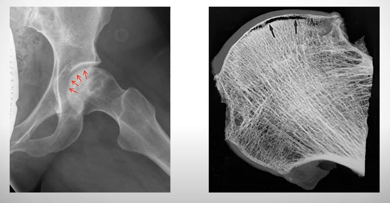
- Densitometry. Required to determine bone density and strength.
If the patient is unable to sit or stand and the pain relief is useless, they should be hospitalized immediately for further surgical treatment.
When to see a doctor urgently
- When there is sharp pain in the movement of the hip joint.
- If the affected leg cannot be supported.
- Detection of edema of the lumbar and femoral region.
- Redness or bruising in the affected area.
There are folk methods for relieving pain in the pelvic joint. It is not worth relying on these tips for a quick recovery. Without a thorough diagnosis, it is impossible to determine the cause of arthralgia and self-medication leads to complications.